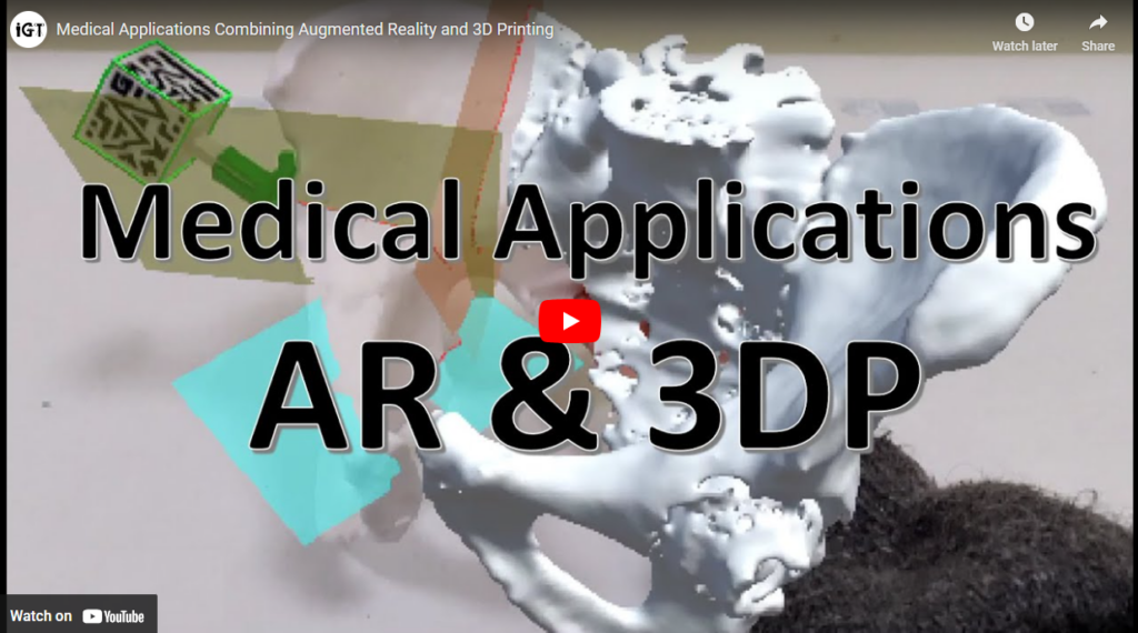
Augmented Reality (AR) superimposes 3D virtual objects to physical objects in real space. In other words, it can provide real-time virtual information directly in our environment. This tool has been lately adopted in many medical areas with exciting benefits. In this group, we also believe in the potential of AR, and so, we have developed several AR applications applied to the clinical field. Our research has been done in collaboration with Hospital Gregorio Marañón and combines this technology with 3D printing.
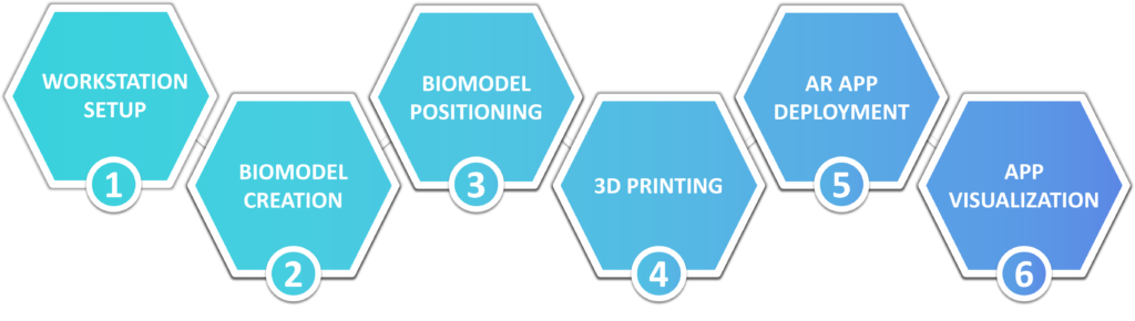
How does it work? We create 3D virtual models of patients’ internal structures using their medical images. On the other hand, we design patient-specific 3D printed surgical guides that can fit into a specific region of the patient’s anatomy. Attached to these guides, we include a 3D printed reference that the AR system can recognize. As a result, we can project the patients’ inside over themselves to create the illusion that we are looking through their skin. With that, we attempt to facilitate surgical planning, assist during patient communication, and enhance advanced surgeries.

HoloLens 2
2026 – Universal AR app for HoloLens 2
AR offers significant potential to enhance surgical precision, improve clinical training, and support intraoperative decision-making. However, developing personalized AR applications for head-mounted displays such as Microsoft HoloLens 2 typically requires advanced technical expertise. Additionally, identifying a robust and accessible method for registering virtual models to the real world remains an open challenge. These limitations hinder the widespread adoption of AR in healthcare, particularly among non-technical clinical users.
This work presents EZ-AR, a user-friendly AR application for Microsoft HoloLens 2 that automates complex tasks such as configuration and deployment, while ensuring low latency, high reliability, and smooth interaction. Ultimately, the system enables non-expert users to independently load and visualize personalized 3D models anchored to physical reference markers. The application accesses files from Google Drive in seconds and displays them in the physical environment using one of two integrated tracking methods: Vuforia or QR code detection. Once configured, the application no longer requires Internet access.
The system’s usability was assessed in terms of detection range and AR projection accuracy under varying surface inclinations and viewing angles. Results show a mean projection error of 3.3 ± 2.2 mm with QR code tracking and 3.9 ± 2.7 mm with Vuforia, both measured at a working distance compatible with typical surgical ergonomics. Further analysis revealed that accuracy is more influenced by viewing angle and surface inclination than by the tracking method itself. Overall, EZ-AR provides a robust and accessible AR framework to democratize AR adoption in clinical environments.
By clicking in the following image you will find some instructions and details on how to use the app as well as a video with sample use cases, although… the limit is your imagination!

2025 – Craniosynostosis surgery
Craniosynostosis is a congenital condition characterized by the premature fusion of cranial sutures, leading to potential complications such as abnormal skull growth, increased intracranial pressure, and cognitive delays. Traditionally, open cranial vault reconstruction (OCVR) has been used to treat this condition. However, it is highly subjective and greatly dependent on the surgeon’s expertise, which can lead to residual deformities and the need for reoperation. More information regarding this disease and its traditional treatment is available at Craniosynostosis Surgery.
Effective preoperative planning can greatly improve surgical outcomes, although the major challenge is accurately translating this plan into the clinical setting. Recently, augmented reality (AR) and 3D printing have emerged as promising technologies to facilitate this endeavor. In this work, we propose three alternatives, leveraging these technologies, to guide the precise repositioning of remodeled bone fragments in the patient.
The three guidance methods are AR on a tablet, AR with Microsoft HoloLens 2, and 3D-printed spacers. The accuracy of each method was assessed by measuring the deviation of each bone fragment from the virtual surgical plan (VSP) in a simulated environment using 3D-printed phantoms based on a 14-month-old boy with trigonocephaly. The same assessment was also performed during his actual surgery.
All three guidance methods demonstrated similar levels of accuracy, with mean placement errors below 1 mm in all cases. The AR systems allowed for real-time adjustments, enhancing precision. Statistical analysis showed no significant differences in error rates between the different methods or attempts.
In conclusion, integrating AR and 3D printing into craniosynostosis surgery holds great potential for improving OCVR. While 3D-printed spacers are useful when digital technologies are unavailable, AR-based methods provide more comprehensive guidance. Nevertheless, our study suggests that the choice may depend more on the specific clinical context, user-specific skills, and available resources rather than on a clear superiority of one method over the others.
The following two videos demonstrates the surgical experience with all devices, as well as the evaluation workflow.


- Pose-Díez-de-la-Lastra A, García-Sevilla M, Tapp A, Tousidonis M, Darriba-Alles JV, Linguraru MG, Pascau J, Ochandiano S. Microsoft HoloLens 2 vs. Tablet-based Augmented Reality and 3D printing for fronto-orbital reconstruction of craniosynostosis: A case study. 3D Printing in Medicine 11, 13 (2025). [doi] – Impact Factor: 3.2 (Q1)
2023 – HoloLens 2 + 3D Slicer
At Universidad Carlos III de Madrid, we are mainly focused on Microsoft HoloLens 2. To date, there has been a lack of software infrastructure to connect 3D Slicer to any augmented reality (AR) device. Our most recent project presents a novel connection approach between 3D Slicer and Microsoft HoloLens 2 using OpenIGTLink. This project has been developed in collaboration with Perk Lab from Queen’s University. The solution is implemented in a 3 elements system: A Microsoft HoloLens 2 headset, the Unity software, and the 3D Slicer platform.
Specifically, we developed an application that transfers geometrical transform and image messages between the platforms. It sends the position of a plane from HoloLens 2 to 3D Slicer. The computer software reslices the CT volume of the patient at this specific pose and sends it back to the AR glasses. This interaction is performed in real-time, so that the user can directly visualize CT reslices of a patient overlayed to the real world. Below you can see a demonstration for pedicle screw placement planning.

All our work is publicly available in this GitHub repository. This repository contains all the resources and code needed to replicate our work on a new computer.
- Pose-Díez-de-la-Lastra A, Ungi T, Morton D, Fichtinger G, Pascau J. Real-time integration between Microsoft HoloLens 2 and 3D Slicer with demonstration in pedicle screw placement planning. Int J CARS (2023). [doi] [pdf, CC BY License] – Impact Factor: 3.421 (Q2)
2022 – Orthopedic oncological surgeries
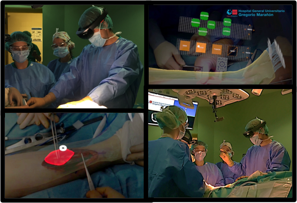
We wanted to go a step forward from the previously used Smartphones and HoloLens 1. Our first task was to evaluate their tracking accuracy and ergonomics compared to HoloLens 1 during experimental and surgical scenarios. Check our free-access paper below to find out our results.
The top left corner shows an external view of the surgical field
- A. Pose-Díez-de-la-Lastra, R. Moreta-Martinez, M. García-Sevilla, D. García-Mato, J. A. Calvo, L. Mediavilla-Santos, R. Pérez-Mañanes, F. von Haxthausen, and J. Pascau. HoloLens 1 vs. HoloLens 2: Improvements in the New Model for Orthopedic Oncological Interventions. Sensors 2022, 22, 4915. [doi] [Open Access under the Creative Commons Attribution-NonCommercial-NoDerivs License]
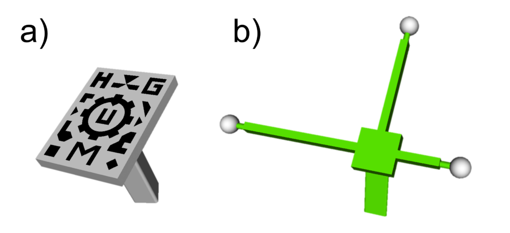
In addition, we have analyzed the HoloLens 2’s tracking performance using two different types of AR markers: a pattern-based and a spheres-based marker. The first recognition system is based on pattern identification using Vuforia. This system uses computer vision algorithms to identify visual patterns in an RGB image acquired with the HoloLens 2’s camera. The second system uses HMD’s RGB and depth camera to detect another AR marker made of three retro-reflective spheres. We tested both strategies in an experimental scenario and during real surgery. Our results were presented at Computer Assisted Radiology and Surgery (CARS) congress, celebrated in Tokyo during June 2022.
- A. Pose-Díez-de-la-Lastra, R. Moreta-Martinez, F. con Haxthausen, M. García-Sevilla, L. Hernández-Álvarez, J. A. Calvo, R. Pérez-Mañanes, and J. Pascau. Analysis of tracking performance with two different HoloLens 2 augmented reality markers for orthopedic oncological surgeries. In: CARS 2022 – Computer Assisted Radiology and Surgery Proceedings of the 36th International Congress and Exhibition, Tokyo, Japan, June 7-11, 2022. Int J CARS 17, 1-147 (2022). [doi]
HoloLens 1
2018 – Orthopedic oncological surgeries
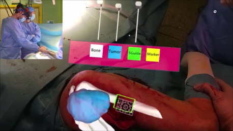
Augmented reality can be an interesting technology for clinical scenarios as an alternative to conventional surgical navigation. However, the registration between augmented data and real-world spaces is a limiting factor. In this work, we propose a method based on desktop 3D printing to create patient-specific tools containing a visual pattern that enables automatic registration. This specific tool fits the patient only in the location it was designed for, avoiding placement errors. This solution has been developed as a software application running on Microsoft HoloLens.
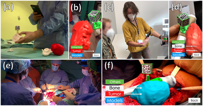
The workflow was validated during the surgical intervention of a patient presenting an extraosseous Ewing’s sarcoma. The application allowed physicians to visualize the skin, bone, and tumor location overlaid on the patient.
- R. Moreta-Martinez, D. García-Mato, M. García-Sevilla, R. Pérez-Mañanes, J. A. Calvo, and J. Pascau. Augmented reality in computer-assisted interventions based on patient-specific 3D printed reference. Healthcare Technology Letters, 1–5 (2018). [doi] [UC3M Research Portal] [pdf, Open Access under the Creative Commons Attribution-NonCommercial-NoDerivs License]
Smartphones and Tablets
2024 – Microtia reconstruction
In addition to orthopedic oncology, the biomedical engineers from the IGT group have also joined efforts with Hospital General Universitario Gregorio Marañón to treat microtia. This is an congenital malformation in which the ears develop incorrectly, resulting in tiny or even non-existent ears. This condition, which affects 1 in every 5,000 births, can not only compromise the patient’s hearing ability but also interfere with everyday situations such as wearing glasses.

The affected ear can be reconstructed during surgery using the patient’s own costal cartilage. Traditionally, the contralateral healthy ear has been used as a template to trace the ideal shape of the new ear in the extracted cartilage, while anatomical measurements usually guide its placement on the patient’s head to ensure symmetrical outcomes. However, this technique is time-consuming, subjective, and highly dependent on the surgeon’s expertise, making it susceptible to shape errors and misplacement.
We present an innovative clinical workflow that combines 3D printing and augmented reality (AR) to increase the objectivity and reproducibility of these procedures. Specifically, we introduce patient-specific 3D cutting templates and remodeling molds to carve and construct the cartilaginous framework that will form the new ear. Moreover, we developed an in-house AR application compatible with any commercial Android tablet. It precisely guides the positioning of the new ear during surgery, ensuring symmetrical alignment with the healthy one and avoiding time-consuming intraoperative linear or angular measurements. Our solution was evaluated in one case, first with controlled experiments in a simulation scenario and finally during surgery.

- Alberto Díez-Montiel, Alicia Pose-Díez-de-la-Lastra, Alba González-Álvarez, José-Ignacio Salmerón, Javier Pascau, Santiago Ochandiano. Tablet-based Augmented reality and 3D printed templates in fully guided Microtia Reconstruction: a clinical workflow. 3D Printing in Medicine 10, 17 (2024). [doi] [pdf, Open Access under the Creative Commons Attribution 4.0 International License]
2020 – DIY app tutorial
At Universidad Carlos III de Madrid and Hospital General Universitario Gregorio Marañón, we have implemented a protocol to develop your own smartphone app combining augmented reality and 3D printing for its use in the medical field. The method describes detailed steps to go from the patient medical image to the development of a smartphone app using free and multi-platform software. This protocol is expected to accelerate the adoption of AR and 3DP technologies by medical professionals. No prior knowledge is required.
Check out the protocol following this link: https://www.jove.com/v/60618/ (Journal of Visualized Experiments)
It will take you to a step-by-step guide, including open access to all the material.
In the following video, we show you some of its applications:
- R. Moreta-Martinez, D. García-Mato, M. García-Sevilla, R. Pérez-Mañanes, J. A. Calvo-Haro, J. Pascau. Combining Augmented Reality and 3D Printing to Display Patient Models on a Smartphone. J. Vis. Exp., 155, e60618 (2020). [doi] [UC3M Research Portal] [pdf, Open Access under the Creative Commons Attribution-NonCommercial-NoDerivs License]
2021 – Orthopedic oncological surgeries
The standard treatment of bone and soft tissue tumors includes complete surgical resection. This procedure still depends on the surgeon’s previous experience and subjective judgment to achieve complete tumor removal, ensuring a safety margin of healthy tissue. For this reason, it is essential to efficiently plan the surgical approach preoperatively to improve surgical outcomes, leave enough surgical margin, and reduce the risk of local recurrence or metastasis
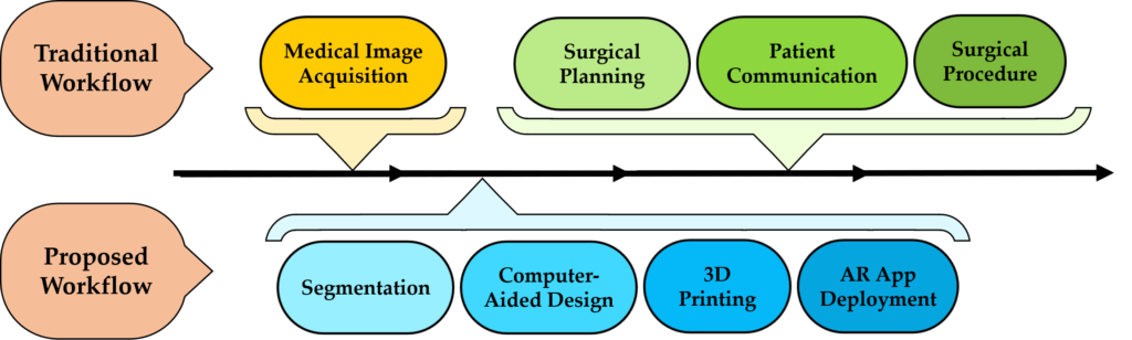
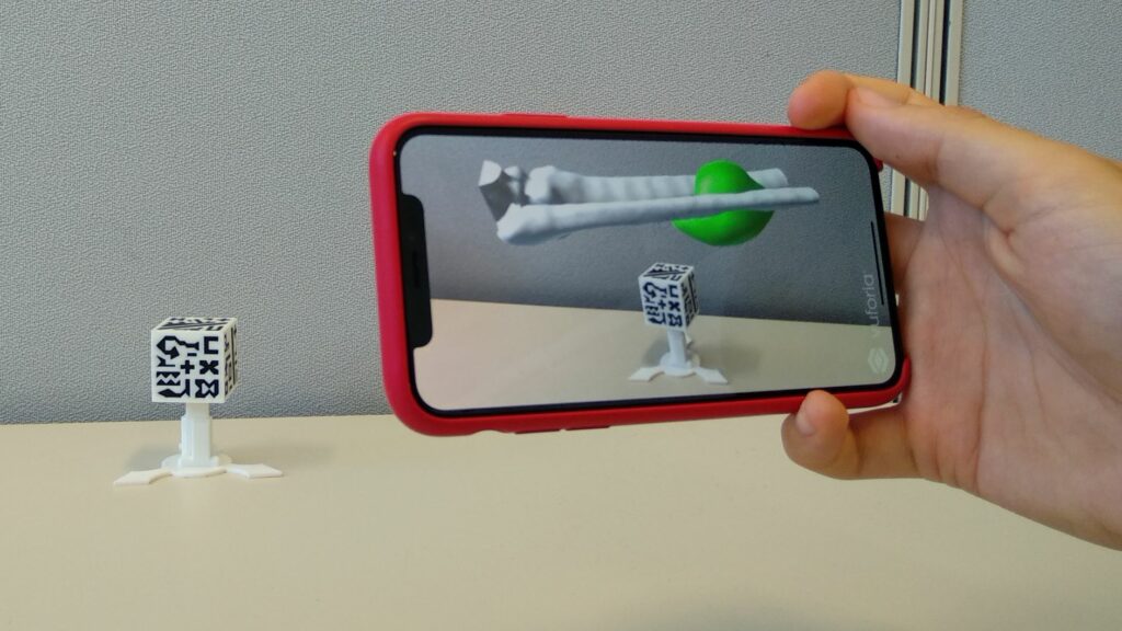
In this project, we propose a new surgical framework that combines augmented reality and 3D printing for their integration in orthopedic oncological surgeries. It includes 3D-printed patient-specific models and tools and a novel smartphone-based AR application. This app displays the internal anatomical structures of the patient in real-time. The system is part of a new surgical workflow in which both technologies assist in preoperative planning, patient communication, and surgical intervention.
- R. Moreta-Martinez, A. Pose-Díez-de-la-Lastra, J. A. Calvo-Haro, L. Mediavilla-Santos, R. Pérez-Mañanes, and J. Pascau. Combining Augmented Reality and 3D Printing to Improve Surgical Workflows in Orthopedic Oncology: Smartphone Application and Clinical Evaluation. Sensors, 21(4), 1370 (2021). [doi] [pdf, Open Access under the Creative Commons Attribution 4.0 International License]
Other Applications
2021 – AR in Craniosynostosis
Related publications
- Pose-Díez-de-la-Lastra A, García-Sevilla M, Tapp A, Tousidonis M, Darriba-Alles JV, Linguraru MG, Pascau J, Ochandiano S. Microsoft HoloLens 2 vs. Tablet-based Augmented Reality and 3D printing for fronto-orbital reconstruction of craniosynostosis: A case study. 3D Printing in Medicine 11, 13 (2025). [doi] – Impact Factor: 3.2 (Q1)
- Alberto Díez-Montiel, Alicia Pose-Díez-de-la-Lastra, Alba González-Álvarez, José-Ignacio Salmerón, Javier Pascau, Santiago Ochandiano. Tablet-based Augmented reality and 3D printed templates in fully guided Microtia Reconstruction: a clinical workflow. 3D Printing in Medicine 10, 17 (2024). [doi] [pdf, Open Access under the Creative Commons Attribution 4.0 International License]– Impact Factor: 3.7 (Q1)
- Pose-Díez-de-la-Lastra A, Ungi T, Morton D, Fichtinger G, Pascau J. Real-time integration between Microsoft HoloLens 2 and 3D Slicer with demonstration in pedicle screw placement planning. Int J CARS (2023). [doi] [pdf, CC BY License] – Impact Factor: 3.421 (Q2)
- Pose-Díez-de-la-Lastra A, Moreta-Martinez R, García-Sevilla M, García-Mato D, Calvo-Haro JA, Mediavilla-Santos L, Pérez-Mañanes R, von Haxthausen F, and Pascau J. HoloLens 1 vs. HoloLens 2: Improvements in the New Model for Orthopedic Oncological Interventions. Sensors, 22(13), 4915 (2022). [doi] [Open Access under the Creative Commons Attribution 4.0 International License] – Impact Factor: 3.576 (Q1).
- R. Moreta-Martinez, D. García-Mato, M. García-Sevilla, R. Pérez-Mañanes, J. A. Calvo-Haro, J. Pascau. Combining Augmented Reality and 3D Printing to Display Patient Models on a Smartphone. J. Vis. Exp., 155, e60618 (2020). [doi] [UC3M Research Portal] [pdf, Open Access under the Creative Commons Attribution-NonCommercial-NoDerivs License]
- R. Moreta-Martinez, A. Pose-Díez-de-la-Lastra, J. A. Calvo-Haro, L. Mediavilla-Santos, R. Pérez-Mañanes, and J. Pascau. Combining Augmented Reality and 3D Printing to Improve Surgical Workflows in Orthopedic Oncology: Smartphone Application and Clinical Evaluation. Sensors, 21(4), 1370 (2021). [doi] [pdf, Open Access under the Creative Commons Attribution 4.0 International License]
- R. Moreta-Martinez, D. García-Mato, M. García-Sevilla, R. Pérez-Mañanes, J. A. Calvo, and J. Pascau. Augmented reality in computer-assisted interventions based on patient-specific 3D printed reference. Healthcare Technology Letters, 1–5 (2018). [doi] [UC3M Research Portal] [pdf, Open Access under the Creative Commons Attribution-NonCommercial-NoDerivs License]
- D. García-Mato, R. Moreta-Martinez, M. García-Sevilla, S. Ochandiano, R. García-Leal, R. Pérez-Mañanes, J. A. Calvo-Haro, J.I. Salmerón, J. Pascau. Augmented reality visualization for craniosynostosis surgery. Comput. Methods Biomech. Biomed. Eng. Imaging Vis., 1–8 (2020). [doi]