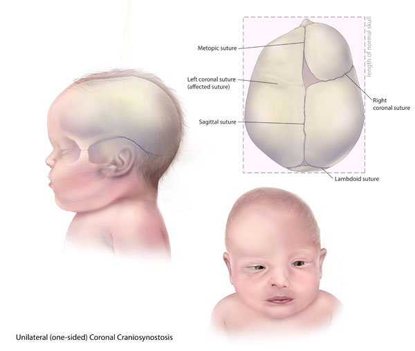
Craniosynostosis is a congenital defect defined as the premature fusion of one or more cranial sutures. This fusion leads to growth restriction and deformation of the cranium, caused by compensatory expansion parallel to the fused sutures. Surgical correction is the preferred treatment in most cases to excise the fused sutures and to normalize cranial shape. Although multiple technological advancements have arisen in the surgical management of craniosynostosis, interventional planning and surgical correction are still highly dependent on the subjective assessment and artistic judgment of craniofacial surgeons. Therefore, there is a high variability in individual surgeon performance and, thus, in the surgical outcomes.
Researchers of our group have worked in collaboration with craniofacial surgeons at Hospital General Universitario Gregorio Marañón (Madrid, Spain) to improve the surgical management of craniosynostosis by reducing subjectivity in all stages of the process, from the preoperative virtual planning phase to the intraoperative performance.
Intraoperative 3D Photography
We have demonstrated that 3D photography is a valuable alternative to CT imaging for the intraoperative evaluation of surgical outcomes during craniosynostosis surgeries. This technology enables accurate 3D reconstruction and cranial shape quantification during the surgical intervention, which can be used to provide intraoperative feedback to surgeons.
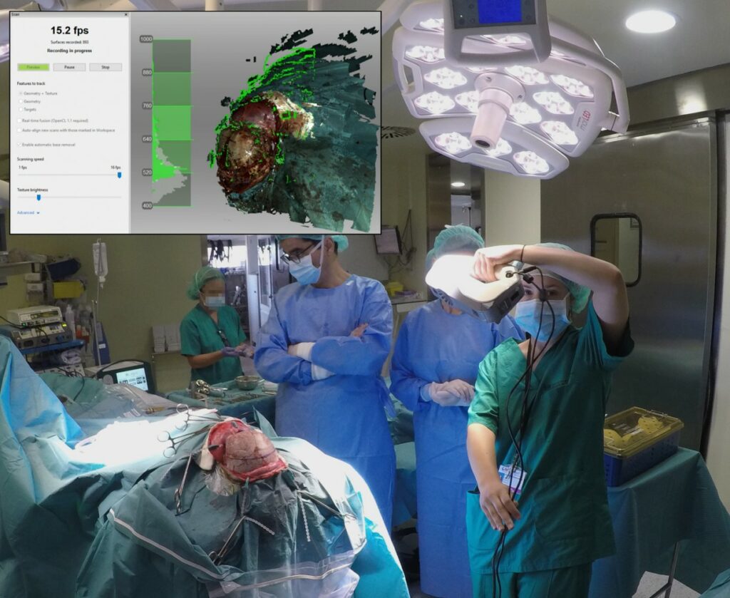
Image-Guided Surgery
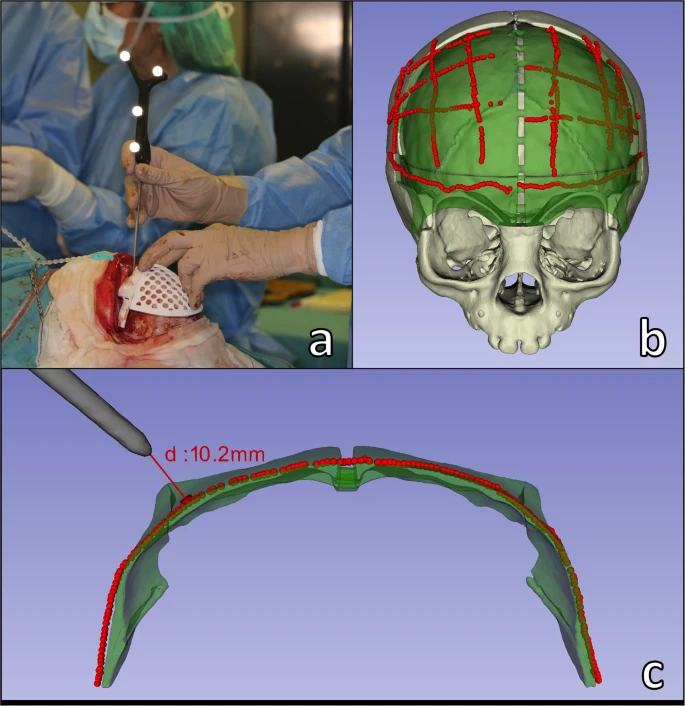
An intraoperative navigation system was developed to provide real-time guidance during open cranial vault remodeling. This system was specifically designed for craniosynostosis surgery, enabling accurate navigation while avoiding the invasive attachment of dynamic reference frames or infant’s head immobilization. The evaluation of the system demonstrated an optimal accuracy for bone fragment positioning, and feasibility of integration into the surgical workflow without substantially increasing operative time.
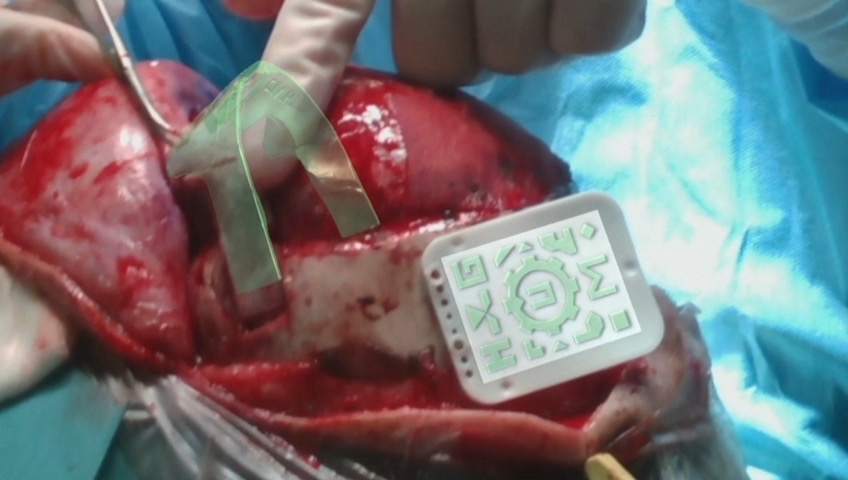
Augmented reality
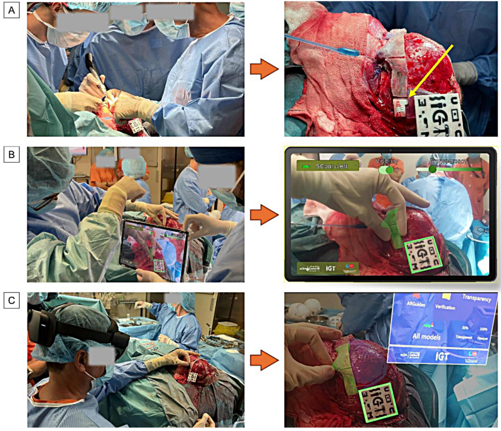
In 2025, we proposed and evaluated three different alternative methods to guide the precise repositioning of remodeled bone fragments in the patient in order to facilitate the translation of the preoperative plan into the clinical setting. The three guidance methods were AR on a tablet, AR with Microsoft HoloLens 2, and the use of 3D printed guides (spacers). More information regarding this work is provided in AR in Craniosynostosis Surgery.
recordings of the procedure are available in Supplementary Video S1 at https://zenodo.org/records/14697459.
In a previous work from 2020, we also proposed and evaluated a workflow for AR visualization to guide the osteotomy and remodeling phases of FOA by overlaying virtual information directly on the surgical field. Our results demonstrated the accuracy of AR technology for surgical guidance in craniosynostosis, and its feasibility for integration into the surgical workflow. The developed software application can be used for visualization in smartphones, head-mounted displays, and external screens.
Related publications
- A. Pose-Díez-de-la-Lastra, M. García-Sevilla, A. Tapp, M. Tousidonis, J.V. Darriba-Alles, M.G. Linguraru, J. Pascau, S. Ochandiano. MicrosoftHoloLens 2 vs. Tablet-based Augmented Reality and 3D printingfor fronto-orbital reconstruction of craniosynostosis: A case study. 3D Printing in Medicine 11, 13 (2025). [doi] – Impact Factor: 3.2 (Q1)
- A. Pose-Díez-de-la-Lastra, L. García-Duarte Sáenz, D. García-Mato, L. Hernández-Álvarez, S. Ochandiano and J. Pascau. Real-Time Tool Detection for Workflow Identification in Open Cranial Vault Remodeling. Entropy 23(7), 817 (2021). [doi] [pdf, CC BY License] – Impact factor: 2.494 (Q2)
- D. García-Mato, M. García-Sevilla, A.R. Porras, S. Ochandiano, J.V. Darriba-Allés, R. García-Leal, J.I. Salmerón, M.G. Linguraru, and J. Pascau. Three-Dimensional Photography for Intraoperative Morphometric Analysis in Metopic Craniosynostosis Surgery. Int J CARS 16, 277–287 (2021). [doi]
- D. García-Mato, R. Moreta-Martinez, M. García-Sevilla, S. Ochandiano, R. García-Leal, R. Pérez-Mañanes, J. A. Calvo-Haro, J.I. Salmerón, J. Pascau. Augmented reality visualization for craniosynostosis surgery. Comput. Methods Biomech. Biomed. Eng. Imaging Vis., 1–8 (2020). [doi]
- D. García-Mato, S. Ochandiano, M. García-Sevilla, C. Navarro-Cuéllar, J. V. Darriba-Allés, R. García-Leal, J. A. Calvo-Haro, R. Pérez-Mañanes, J. I. Salmerón, and J. Pascau. Craniosynostosis surgery: workflow based on virtual surgical planning, intraoperative navigation and 3D printed patient-specific guides and templates. Scientific Reports, 9, 17691 (2019). [doi] [UC3M Research Portal] [pdf, Open Access under the Creative Commons Attribution 4.0 International License]